What the imaging standards are actually asking of you
Strip away the clause numbering and the imaging standards ask five questions. Do you know what services you provide and by whom. Are the people doing the work qualified for it. Is the work done to a written method that reflects what actually happens. Is the equipment safe, maintained and monitored. And can you show that you measure the quality of the service and act on what you find. Every requirement in the chapter is a specific instance of one of those five.
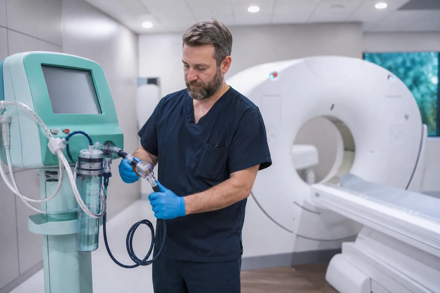
In the current edition the imaging requirements sit within the chapter dealing with access, assessment and continuity of care, alongside the laboratory ones, which occasionally leads departments to assume the two are handled identically. They are not. Imaging carries a radiation safety programme that has no laboratory equivalent, an equipment quality assurance regime tied to AERB obligations, and contrast administration as a clinical procedure with its own consent and reaction management expectations.
The most useful preparation posture is to stop treating this as an accreditation exercise and treat it as an operating manual you happen to be showing someone. Departments that write documents for the assessment produce shelves of unread SOPs and a visible gap between paper and practice, which is precisely what a competent assessor is trained to find. Departments that write down what they actually do, then improve it, walk through the assessment without a scramble.

The scope of services document that everything else hangs from
The scope of services is a short document and it governs more than its length suggests. It states which modalities you provide, which examinations within each, the hours during which each is available, which services are outsourced and to whom, and which are explicitly not provided. Everything downstream is judged against it: your staffing, your SOPs, your equipment list, your emergency arrangements and your referral pathways all have to be consistent with what this document claims.
The recurring problem is drift. The scope document says MRI is available from nine to five on weekdays. In practice the department has been running emergency MRI at night for a year because a neurologist asked and it became normal. Neither the staffing plan nor the SOP nor the on-call arrangement reflects it. That is not an administrative untidiness; it is a service running without a defined competence and cover model, which is exactly the risk the standard exists to prevent.
Review it annually with the clinical heads who actually use imaging, not only within radiology. Ask three questions: what have we started doing that is not written here, what is written here that we no longer do, and what do our referrers believe we provide. The third question is the interesting one, because a mismatch between what physicians expect at two in the morning and what the department is staffed for is a patient safety issue long before it is an accreditation finding.
What the scope of services has to state to be usable
- Every modality and examination offered, with hours of availability for each
- Which examinations require prior appointment and which are available on demand
- Services provided by outsourced or visiting arrangements, and by whom
- Examinations explicitly not performed, with the referral route for them
- Emergency and after-hours arrangements including reporting cover
The SOPs an assessor opens first
Assessors do not read your SOP folder end to end. They open a small number of documents that reveal whether the set is genuine, and the choices are predictable. Patient identification before an examination, because misidentification is the highest-frequency serious error in imaging. Contrast administration including screening, consent and reaction management. Radiation safety including pregnancy screening for women of childbearing age. Handling of critical and unexpected findings. And the procedure for reporting, verification and amendment.
The test applied is consistency between the document and the person in front of them. An assessor reads that pregnancy status is confirmed and recorded before any examination involving ionising radiation, then walks to the CT console and asks the technologist to show where that is recorded. If the answer is that it is asked verbally and not documented, the SOP is aspirational. The correction is not to rewrite the SOP; it is to add the field and use it.
Version control quietly fails more departments than content does. An SOP with no issue date, no review date, no author and no evidence that anyone was trained on the current version is treated as uncontrolled regardless of how good the text is. Keep a simple document register showing version, issue date, next review and training record, and keep superseded versions retrievable rather than destroyed, because a question about practice two years ago is a question about the version in force then.
Imaging SOPs to have current, trained and version-controlled
- Patient identification and correct-site verification before examination
- Pregnancy screening and documentation for examinations using ionising radiation
- Contrast screening, consent, administration and reaction management
- Communication and acknowledgement of critical and unexpected findings
- Reporting, verification, amendment and issue of duplicate reports
Choosing imaging quality indicators you will actually use
You are expected to monitor the quality of the imaging service, and you are largely free to choose which indicators do that. The temptation is to adopt a long list because it looks thorough. The result is a department collecting fifteen numbers, analysing none, and unable to explain what any of them changed. Four or five indicators, collected reliably and reviewed monthly with actions recorded, is a stronger position in every respect.
Choose indicators that cover different failure types rather than five variations of the same thing. Report turnaround gives you throughput. Repeat and reject rate gives you technical quality and dose. Adverse contrast reactions give you clinical safety. Discrepancy or peer review findings give you interpretive quality. Downtime of critical equipment gives you availability. Waiting time for an appointment gives you access. Pick from across that spread rather than three timing measures.
Define each one properly before you start collecting, because a redefined indicator destroys its own trend. Write the numerator, the denominator, the inclusions and exclusions, the data source and the person responsible. Where the number is pulled from a system rather than counted by hand, note the report or query that produces it, so the figure can be reproduced by someone else next year. HealUDoc dashboards can hold indicator definitions as versioned configuration, which lets you show an assessor exactly which rule produced a given month's figure.
Equipment, radiation safety and the AERB overlap
The imaging chapter and your AERB obligations cover much of the same ground from different angles, and an efficient department maintains one evidence file rather than two. Equipment inventory, planned preventive maintenance, calibration and quality assurance testing, breakdown records with response times, and validity of licences all serve both purposes. Where the accreditation view adds something is in asking whether the maintenance actually happened on schedule and what you did when it did not.
Personnel monitoring is examined closely because it is easy to verify. An assessor will ask to see the current dose records for radiation workers, ask what your investigation level is, and ask what happened the last time someone exceeded it. A department that produces a folder of unopened reports has monitoring in name only. The expectation is that someone reviews each cycle, follows up missing badges, and escalates anomalies with a written outcome.
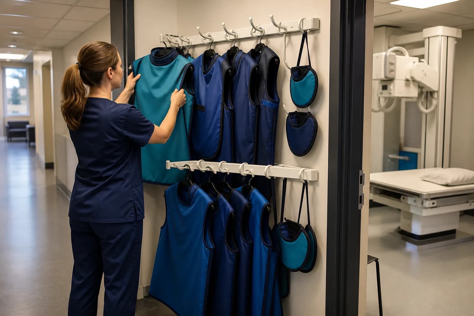
Protective equipment gets checked physically. Lead aprons and thyroid shields have to be present in adequate number, in usable condition, stored on hangers rather than folded, and periodically checked for cracks with the check recorded. This is the single most commonly failed physical item in imaging assessments, and it costs almost nothing to fix. Put a numbered inventory on the wall and a quarterly integrity check against it.
The evidence file, item by item
Assemble the evidence once and maintain it, rather than reconstructing it before each assessment. It should contain the scope of services, the staffing list with qualifications, registrations and privileging, the equipment inventory with licences and quality assurance certificates, the SOP register with training records, the indicator data with monthly analysis and actions, the radiation safety documentation including personnel dose records, and the incident and adverse event records with their closure.
Keep it in the order an assessor works, which is roughly people, then process, then equipment, then outcomes. Being able to hand over a structured file and answer questions from it changes the assessment from an interrogation into a review. Departments that keep everything in individual drives and personal folders spend the assessment fetching things, which reads as poor control whether or not the underlying practice is sound.
One caution about over-preparation. A file that is beautifully assembled and describes a department nobody recognises is worse than a modest file that matches reality, because the mismatch surfaces in the first staff interview. If your indicator data is thin because you started measuring three months ago, say so and show the three months. Honest partial evidence with a plan attracts a far better response than polished evidence that the technologist on duty contradicts.
Documents to keep in the imaging evidence file at all times
- Scope of services, current version, with review date and sign-off
- Staff list with qualifications, council registration numbers and privileging
- Equipment inventory with AERB licence status and quality assurance reports
- SOP register with version, issue date, review date and training attendance
- Twelve months of indicator data with monthly analysis and recorded actions
“The assessor spent ten minutes on our documents and an hour with our technologists. What we had written mattered far less than whether the person at the console described the same process.”
Non-conformities that repeat year after year
The same findings recur across imaging departments with striking consistency. Pregnancy status not documented before examinations using ionising radiation. Lead protective equipment present but with no integrity check record. Critical findings communicated but with no acknowledgement recorded. Indicators collected but never analysed, or analysed with no action recorded. And a scope of services that no longer matches what the department does after hours.
Each of these is cheap to close and none of them closes itself. The pattern in departments that keep repeating them is that the correction is treated as a document change rather than a workflow change. Adding a line to an SOP that pregnancy status will be documented does nothing unless a field exists on the screen the technologist uses and the study cannot proceed without it. Close non-conformities at the point of work.
Run your own internal assessment two or three months before the external one, using someone from outside the department, and score honestly against the same five questions the standards ask. The value is not in the score. It is in finding, while there is still time, that the thing everyone assumed was handled has been handled by one person who left in January. That discovery is worth the discomfort of an internal audit that finds real problems.